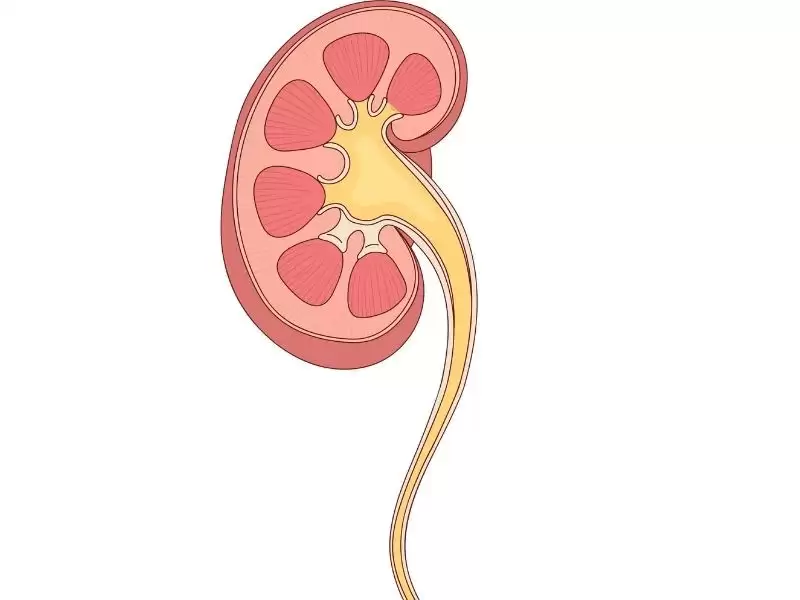
Ureteropelvic Junction Obstruction (UPJ Obstruction) is an important urinary system disorder that occurs due to narrowing at the starting point of the tube (ureter) that carries urine from the kidney to the bladder. This condition prevents urine from draining adequately from the kidney and may gradually cause dilation and damage to kidney tissue. UPJ obstruction, which can occur in both children and adults, is generally congenital but may also develop later in life. If not diagnosed early, it can cause reduced kidney function and serious health problems. Therefore, understanding the causes, symptoms, diagnostic methods, and treatment options is extremely important for protecting kidney health.
- What Is Ureteropelvic Junction Obstruction (UPJ Obstruction)?
- What Causes UPJ Obstruction?
- What Are the Symptoms of UPJ Obstruction?
- What Are the Diagnostic Methods for UPJ Obstruction?
- Treatment of UPJ Obstruction
- Recovery After UPJ Obstruction Surgery
- Frequently Asked Questions About Ureteropelvic Junction Obstruction
- Frequently Asked Questions About Ureteropelvic Junction Obstruction (UPJ Obstruction)

What Is Ureteropelvic Junction Obstruction (UPJ Obstruction)?
Ureteropelvic junction obstruction (UPJ obstruction) is a medical condition in which urine flow is restricted due to narrowing at the point where urine passes from the kidney into the urinary tube (ureter). Also known medically as ureteropelvic junction narrowing, this condition may negatively affect kidney function and eventually lead to dilation of the kidney's collecting system (hydronephrosis). It may be congenital or acquired. Congenital UPJ obstruction generally results from a structural abnormality of the urinary tract and is diagnosed during childhood. However, in some cases, it may not be detected until adulthood. Acquired narrowing usually develops due to kidney stones, infections, surgical interventions, or trauma. Ureteropelvic junction obstruction is particularly important because, if not identified and treated early, it may lead to loss of kidney function.

What Causes UPJ Obstruction?
The causes of UPJ obstruction may be congenital or acquired. Congenital ureteropelvic junction obstruction is often caused by abnormal development of the muscular layer at the point where the ureter connects to the kidney. This prevents urine from flowing normally and gradually increases pressure within the kidney. In some cases, an abnormal blood vessel (accessory vessel) may be present near the junction between the kidney and ureter. This vessel may compress the urinary tract and restrict the passage of urine.
Acquired narrowing usually develops as a result of urinary tract infections, kidney stones, trauma, surgical procedures, or pressure from tumors. In particular, damage to the ureter during the passage of a kidney stone may cause narrowing later on. In some rare cases, fibrosis (thickening of connective tissue) around the ureter may also lead to narrowing at the ureteropelvic junction.
What Are the Symptoms of UPJ Obstruction?
Symptoms of UPJ obstruction may vary depending on the severity and duration of the narrowing. Some patients may remain asymptomatic for a long time, while others may experience noticeable symptoms early on. The most common symptom is pain in the kidney area (the flank or back). This pain is usually intermittent and may become more severe particularly after increased fluid intake or alcohol consumption.
In advanced cases, symptoms such as nausea, vomiting, a feeling of abdominal fullness, and difficulty urinating may occur. Some patients may notice blood in the urine (hematuria). In children, irritability, frequent urination, and abdominal swelling may be observed. One of the most serious complications is impaired kidney function. Long-standing untreated UPJ obstruction may cause permanent kidney damage. Therefore, early diagnosis and treatment are vital.
What Are the Diagnostic Methods for UPJ Obstruction?
In addition to physical examinations and laboratory tests, imaging techniques play a major role in diagnosing UPJ obstruction. Initially, ultrasonography (USG) may detect kidney dilation (hydronephrosis). Ultrasound is particularly preferred in children because it is safe and easy to perform.
Renal scintigraphy (DTPA or MAG-3) may be performed to evaluate kidney function. This examination helps determine whether urine flow is slowed and how well the kidney is functioning. Computed tomography (CT) and magnetic resonance imaging (MR urography) provide detailed information about the precise location and cause of the obstruction.

In some cases, specialized radiological methods such as retrograde pyelography are also used to examine the anatomical structure of the ureter in greater detail. An accurate diagnosis generally requires the combined assessment of multiple tests. This makes it possible to determine the cause and severity of the narrowing, as well as the kidney damage it has caused, and to establish the most appropriate treatment plan.
Treatment of UPJ Obstruction
Treatment of UPJ obstruction is determined according to the severity of the condition, kidney function, and the underlying cause. In mild cases, regular monitoring alone may be sufficient if kidney function is preserved. However, if there is a significant obstruction to urine flow or kidney dilation and loss of function are detected, surgical treatment is necessary.
The most commonly performed surgical procedure is pyeloplasty. During this operation, the narrowed section is removed and a new connection is created to allow urine to flow freely. Today, this operation is performed using laparoscopic or robotic pyeloplasty techniques, which provide a more comfortable and faster recovery compared with open surgery. These methods involve small incisions, and patients are usually discharged from the hospital within a few days.
In some cases, a thin tube called a double-J stent is temporarily inserted to facilitate urine drainage. The stent helps urine pass from the kidney to the bladder, reducing pressure and allowing the kidney to recover.
Recovery After UPJ Obstruction Surgery
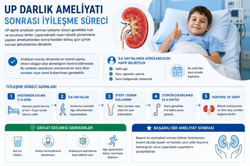
Recovery after UPJ obstruction surgery is generally quick and uncomplicated. Following laparoscopic or robotic procedures, patients may return to their normal activities within a few days. The most important aspect of the postoperative period is checking whether urine is draining properly. Therefore, a urinary catheter or stent may need to be used temporarily after the operation.
During the first weeks after surgery, patients may experience mild pain, burning during urination, or abdominal discomfort. These symptoms usually disappear within a short time. Regular follow-up examinations should be performed as recommended by the doctor, adequate fluids should be consumed, and attention should be paid to signs of infection.
The stent is usually removed after 4–6 weeks. Following this period, kidney function and urine flow are reassessed. After a successful pyeloplasty, kidney function improves significantly, and patients can continue their lives for many years without experiencing problems.
Ureteropelvic junction obstruction (UPJ obstruction) is a condition that can be completely treated when diagnosed early. If symptoms such as pain in the urinary tract, frequent infections, or blood in the urine are noticed, a urology specialist should be consulted without delay. Thanks to modern surgical techniques, UPJ obstruction operations can now be performed safely and effectively. Early intervention can help protect kidney health and improve quality of life.
Frequently Asked Questions About Ureteropelvic Junction Obstruction
Frequently Asked Questions About Ureteropelvic Junction Obstruction (UPJ Obstruction)
Is Ureteropelvic Junction Obstruction (UPJ Obstruction) a Dangerous Condition?
Yes, if left untreated, ureteropelvic junction obstruction can negatively affect kidney function. Prolonged urine accumulation can damage kidney tissue and cause permanent kidney failure. However, with early diagnosis and appropriate treatment, the condition can be brought completely under control.
At What Ages Does Ureteropelvic Junction Obstruction (UPJ Obstruction) Occur?
UPJ obstruction may be congenital or may develop later in life. In newborns and children, it usually develops due to congenital causes, while in adults, it may result from factors such as stones, infections, or trauma.
What Are the Symptoms of Ureteropelvic Junction Obstruction (UPJ Obstruction)?
The most common symptom is flank or back pain felt in the kidney area. Other complaints may include blood in the urine, nausea, vomiting, abdominal swelling, and frequent urination. In some patients, the condition may progress without any symptoms.
Is Surgery for Ureteropelvic Junction Obstruction (UPJ Obstruction) a Difficult Operation?
Thanks to modern laparoscopic and robotic pyeloplasty techniques, operations for UPJ obstruction are quite safe and comfortable. These procedures, performed through small incisions, have a short recovery period and a high success rate.
Can Ureteropelvic Junction Obstruction (UPJ Obstruction) Recur?
The success rate after surgery is very high, and the obstruction generally does not recur. However, in some patients, narrowing may develop again due to factors such as scar tissue formation or infection after surgery. Therefore, regular medical follow-up is important.
When Can Patients Return to Normal Life After Surgery for Ureteropelvic Junction Obstruction (UPJ Obstruction)?
After surgery performed using laparoscopic or robotic techniques, patients are generally discharged within a few days and can return to their normal lives within 1-2 weeks. Avoiding strenuous physical activities and following the doctor's recommendations helps speed up the recovery process.